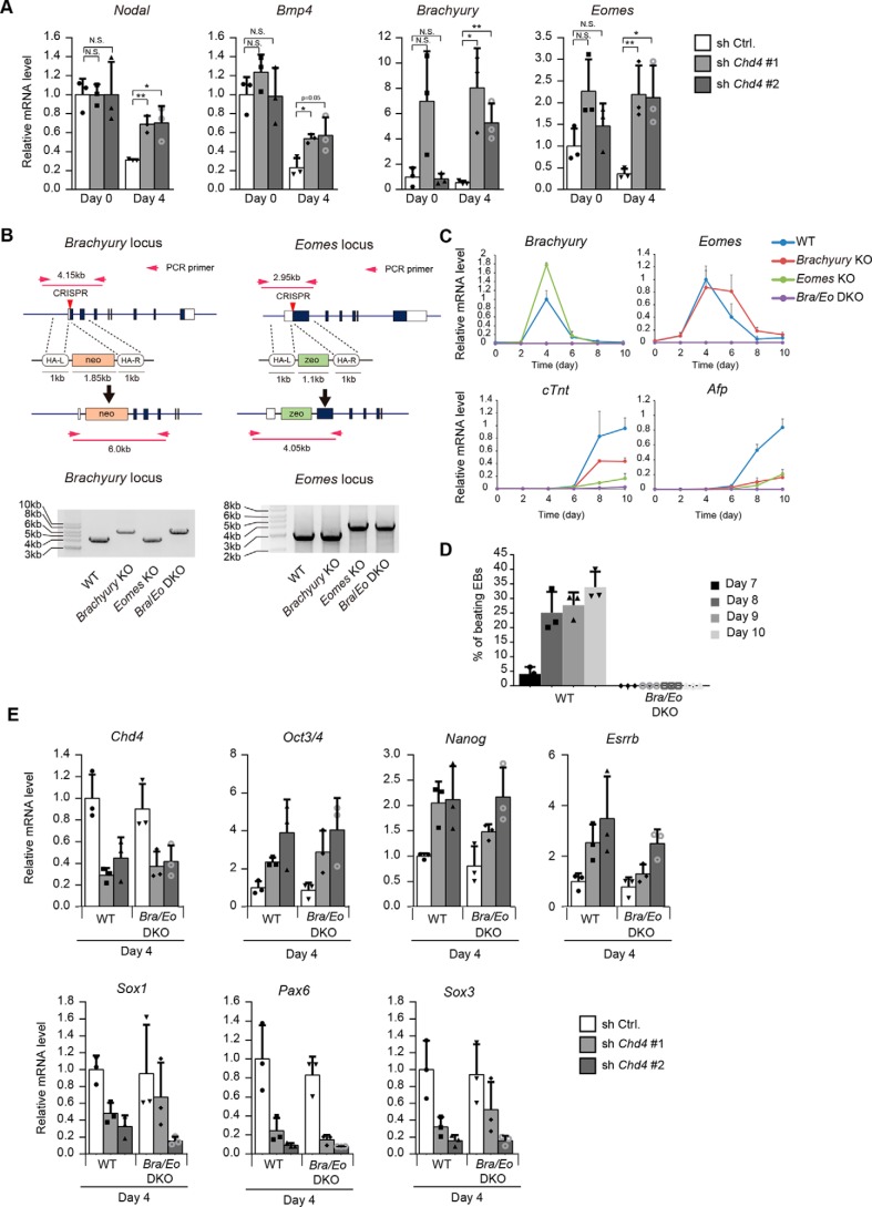
Figure 5.
Inhibition of mesendodermal differentiation does not affect impaired neural differentiation caused by Chd4 knockdown. A, qRT-PCR analysis. Each mRNA level was normalized to the β-actin level, and the value of control shRNA-expressing cells at day 0 was set to 1. B, schematic representation of the strategy to generate Brachyury and Eomes knockout ESCs (left). Proper targeting of Brachuyury and Eomes loci is indicated by PCR genotyping (right). C, qRT-PCR analysis of Brachyury, Eomes, cTnt (mesodermal gene), and Afp (endodermal gene) at the indicated time points of embryoid body formation in WT, Brachyury single knockout, Eomes single knockout, and Bra/Eo DKO cells. The expression levels were normalized to that of β-actin. The y axis values represent the expression levels relative to the maximum expression levels in WT cells. D, percentages of embryoid bodies containing the beating area during the differentiation of WT and mutant cells. E, WT ESCs and Brachyury and Eomes double knockout (Bra/Eo DKO) ESCs were subjected to the experiments shown in Fig. 1A. The mRNA levels at day 4 were determined. Each mRNA level was normalized to the β-actin level, and the value of control shRNA-expressing WT ESCs was set to 1. A and C–E, data are shown as the means ± S.D. (n = 3 independent experiments). A, *, p < 0.05, and **, p < 0.01. N.S., not significant. The p values were calculated using Student's unpaired two-tailed t tests compared with cells on the same day.

