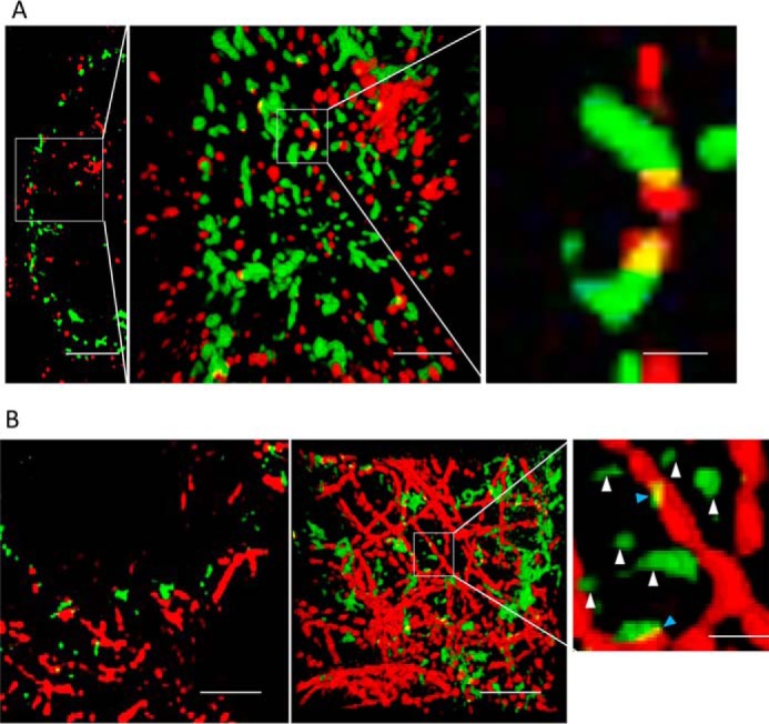
Figure 3.

The majority of polyQ aggregates do not colocalize with HDAC6 or microtubules. A, left panel, a section slice showing cytosolic aggregates (green) in HEK cells that are immunostained against HDAC6 (red). Middle panel, projected view from a 3-D rendering of the data corresponding to the marked region in the left panel. Right panel, magnified view of polyQ aggregate clusters contained in the region indicated in the middle panel to which HDAC6 is bound. Only a small fraction of the clusters colocalize with HDAC6. Scale bars from left to right, 4, 1, and 300 nm, respectively. B, left panel, a section slice of cytosolic aggregates (green) in HEK cells that are immunostained against α-tubulin (red). Middle panel, same field of view, projecting full 3-D data set. Right panel, magnified view of small polyQ clusters localized near microtubules (blue arrowheads). The yellow color indicates areas where particles are in close association. White arrows point to “free” aggregates not associated to microtubules (white arrowheads). Scale bars from left to right, 2, 2, and 400 nm, respectively.
