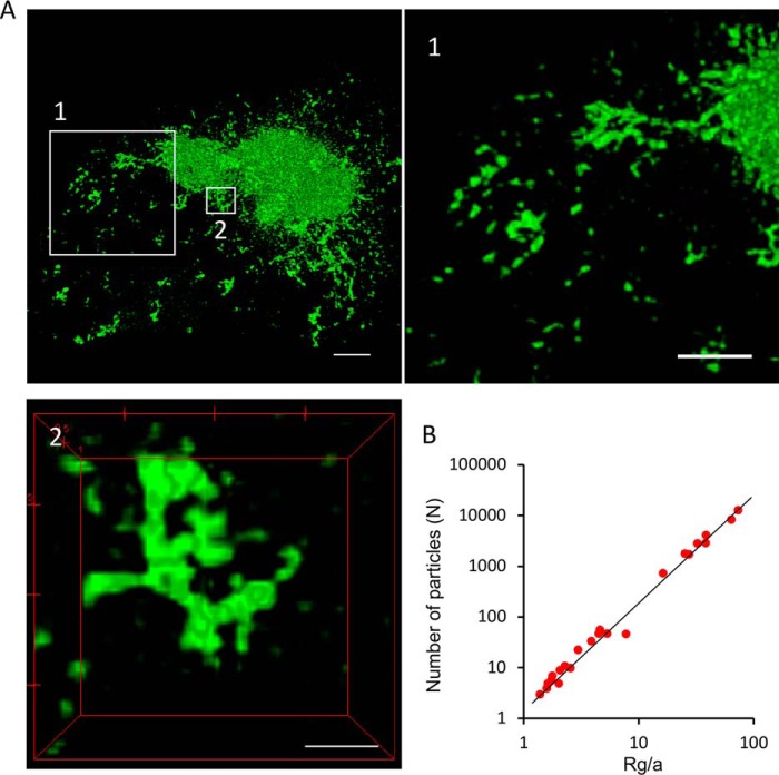
Figure 4.
High-resolution optical imaging of the morphology of an intracellular HDQ72-SNAP aggresome. A, high-resolution SIM images of an aggresome and its peripheral regions. The highlighted regions (boxes 1 and 2) are shown in the corresponding panels. Scale bars, 2 μm. Panel 1, zoomed-in view of peripheral region of the aggresome containing individual, unconnected aggregate fragments that are not associated with the main body of the aggresome. Panel 2, example of amorphous small protrusions from the aggresome surface, containing fused aggregate fragments that have arrived from the cytosol. Scale bars, 1 μm (1) and 500 nm (2). B, number of particles N in an aggregate as a function of cluster radius of gyration Rg for all aggregate clusters, from which the fractal dimension Df is inferred; Rg/a is the gyration radius normalized to the radius of cluster unit. a refers to the radius of an elementary building block made up of small aggregate clusters that, for modeling purposes, is assumed to be spherical.

