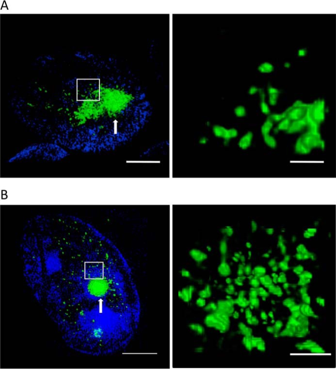
Figure 7.

Intranuclear aggregates in HEK cells have a similar morphology to those observed in an R6/2 mouse model. A, 3-D SIM images of intranuclear EGFP-HDQ72 aggregates (green) in the HEK cell model expressing SNAP-HDQ72. Numerous small clusters and a large aggregate were embedded inside the nucleus. The right-hand panel shows a zoomed-in version of the region in the white rectangle. Scale bars, 2 μm (left panel) and 500 nm (right panel). The cells were stained with Hoechst 33342 (blue). B, 3-D SIM images of immunostained intranuclear polyQ aggregates (green) in the R6/2 mouse model at 14 weeks of age. The aggregates feature a similar morphology to that observed in the cell model. Right panel, zoomed-in region shows the amorphous clusters. Scale bars, 2 μm (left panel) and 500 nm (right panel). The cells were stained with Hoechst 33342 (blue).
