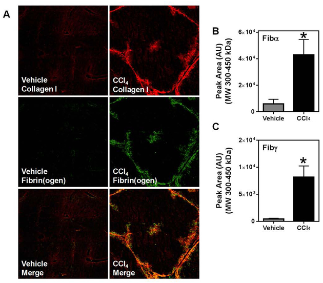
Fig. 1: Deposition of high molecular weight fibrin(ogen) co-localizes with collagen type I following chronic CCl4 exposure.
Wild-type mice were treated with CCl4 or vehicle for 4 weeks, and hepatic fibrin(ogen) and collagen deposition were assessed 3 days after the last injection. (A) Representative photomicrographs (4x virtual objective) of immunofluorescent labeling of collagen type I, fibrinogen, and image overlay in liver sections. HMW cross-linked Fibα (B) and Fibγ (C) detected in liver tissues. Data are presented as mean + SEM (n=6 mice per group). * p < 0.05 compared to vehicle-treated mice.

