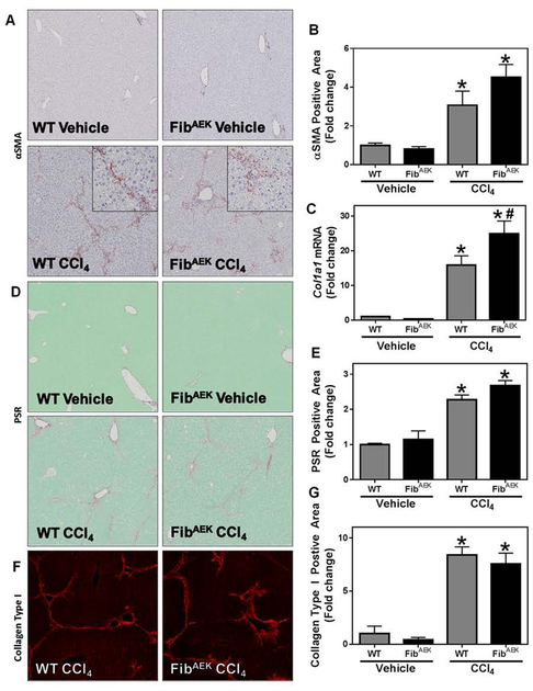
Fig. 3: Effect of FibAEK expression on CCl4-induced experimental hepatic fibrosis.
Wild-type and FibAEK mice were treated with CCl4 or vehicle for 4 weeks and hepatic fibrosis was assessed 3 days after the last injection. (A) Representative photomicrographs (4x virtual objective, 20x insert) of hepatic αSMA labeling. (C) Hepatic Col1a1 mRNA expression. (D) Representative photomicrographs (4x virtual objective) of hepatic PSR staining. (F) Representative photomicrographs (4x virtual objective) of hepatic collagen type I labeling. (B, E, G) Quantification of positive staining area. Data are presented as mean + SEM (n=5–11 mice per group). * p < 0.05 compared to vehicle-treated mice, # p < 0.05 compared to wild-type mice of same treatment group.

