Figure 1.

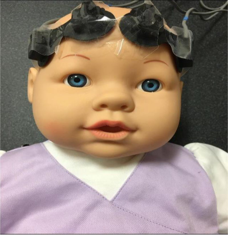
Placement of NIRS optodes in the frontotemporal region on both cerebral hemispheres is shown on a mannequin. In our clinical studies, we wrap the optodes using co-band and ensure that the optodes are firmly attached to the scalp.

Placement of NIRS optodes in the frontotemporal region on both cerebral hemispheres is shown on a mannequin. In our clinical studies, we wrap the optodes using co-band and ensure that the optodes are firmly attached to the scalp.