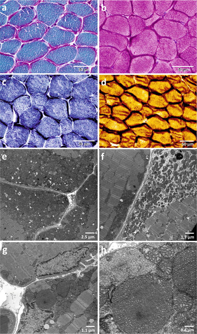
Fig. 1.
Light and electron microscopy of the patient muscle. The muscle biopsy specimen was obtained at the age of 16 years. The muscle shows a massive presence of ragged-red fibers, which are lined by subsarcolemmal mitochondrial accumulations, which impose purple in the Gömöri trichrome stain (a), dark red in the haematoxylin eosin (HE) stain (b), dark blue in the succinate dehydrogenase (SDH) stain (c), and dark brown in the cytochrome c oxidase (COX) stain (d). Electron microscopic images show massive accumulations of mitochondria below the sarcolemma (e) and between the myofibrils (f); presence of giant mitochondria in the muscle measuring up to 3 μm in diameter (g, h)

