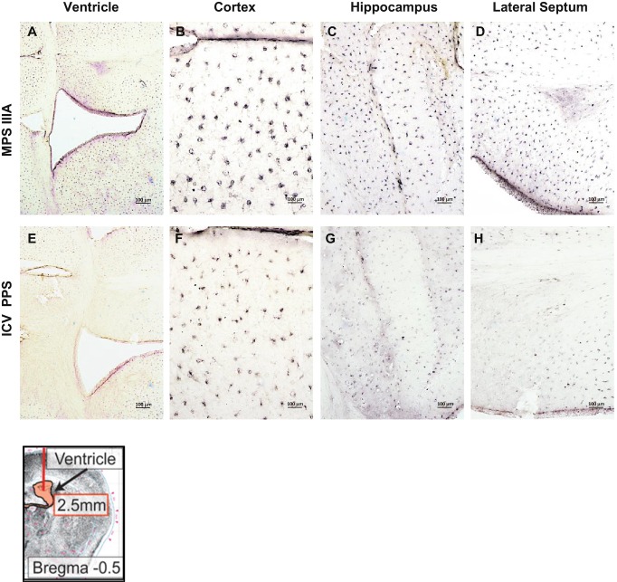
Fig. 8.
IL-B4 staining in MPS IIIA mice treated with PPS by ICV infusion. Panels (a–d) show representative images from 16-week-old, sham treated MPS IIIA mice (ventricle, cortex, hippocampus and lateral septum, respectively). Panels (e–h) show representative images from MPS IIIA mice of the same age that received ICV PPS infusions. A schematic showing the site of injection (arrow) is provided in the lower left

