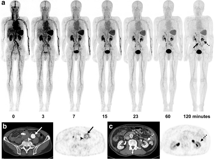
Fig. 2.
18F-GP1 PET/CT and CT images of a 73-year-old man who had undergone endovascular abdominal aortic aneurysm repair surgery. Anterior maximum intensity projections of 18F-GP1 PET/CT show positive 18F-GP1 accumulation in the inner surface of abdominal aortic graft (arrow) and the lower portion of the left kidney (dotted arrow), which are increasingly distinct in later images as background 18F-GP1 activity clears over time via urinary and hepatobiliary excretion (a). Transaxial CT and PET images show increased 18F-GP1 uptake in the inner surface of the left iliac artery graft (b, arrows), but no 18F-GP1 uptake in the chronic intraluminal thrombus in the right iliac aneurysmal sac (b, arrow heads). Additional positive 18F-GP1 uptake is observed in the non-enhancing renal ischaemic area involving the lower portion of the left kidney (c, dotted arrows)

