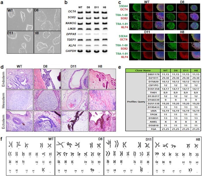
Fig. 3. Characterization of HLA-B–engineered iPSC clones.
a Representative morphologies of HLA-B-engineered iPSC clones. Images were acquired on a Leica microscope (Leica Microsystems Ltd, EC3, image 40 × ). Expression analysis of stemness markers in the wild-type and HLA-B-engineered iPSC clones was performed at the b mRNA and c protein levels by RT-PCR and immunofluorescence staining, respectively (scale bar, 200 μm). d Teratoma assay with wild-type and HLA-B-engineered iPSCs (scale bar, 200 μm). e Genetic profiling of wild-type and HLA-B-engineered iPSC clones. STR was based on the multiplex analysis of 15 loci and the amelogenin gender-determination marker. f Representative karyotypes of wild-type and HLA-B-engineered iPSC clones

