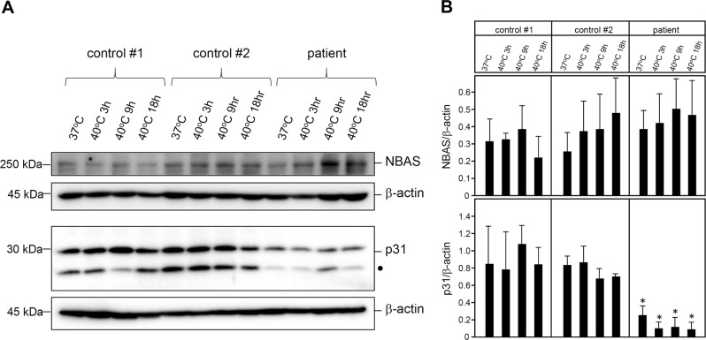
Fig. 5. Western blot of NBAS and p31 in cultured fibroblasts from the patient.
a NBAS protein levels were not decreased even after a shift in the culturing temperature from 37 to 40 °C; however, p31 protein levels were significantly decreased compared to the control subjects. β-actin was used as a loading control. The 15-kDa bands in the p31 western blot indicated by filled circles were isoforms of p31. A typical example of three repeated experiments is shown. b Quantification of NBAS and p31 protein levels. In each case, n = 3, and the mean ± standard deviation (SD) is shown. * denotes P < 0.01 compared to the controls

