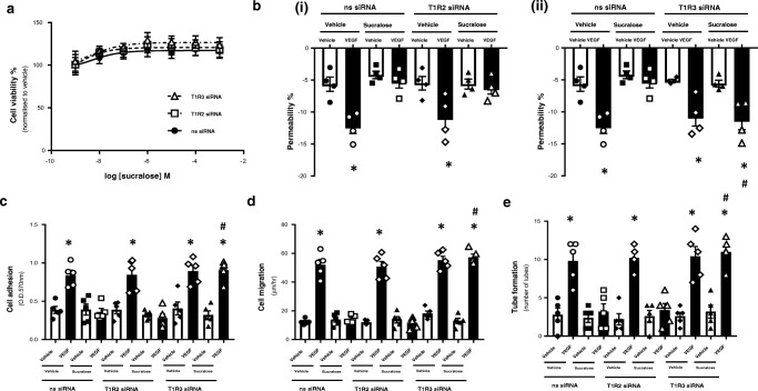
Fig. 4.
Inhibition of T1R3, but not T1R2, blocks the protective effect of sucralose on VEGF-induced angiogenesis in the retinal microvascular endothelium. RMVEC were transiently transfected with T1R2 or T1R3 siRNA, or a non-specific scrambled siRNA (ns) for 24 h prior to treatment with sucralose (0.1 mM) and VEGF (100 ng/ml). Panel a: cell viability of RMVEC was measured by CCK8 assay following exposure to sucralose (1 nM–1 mM). Panel b: changes in retinal endothelial cell monolayer permeability were determined using the FITC-dextran permeability assay in RMVEC with T1R2 (i) or T1R3 (ii) siRNA knockdown. Panel c–e: changes in retinal endothelial cell: adhesion (panel c), migration (panel d), tube formation (panel e) were measured following exposure to sucralose (0.1 mM) in the presence (closed bars) and absence (open bars) of VEGF (100 ng/ml). n = 5. Data is expressed as mean ± S.E.M. *p < 0.05 versus vehicle for VEGF, #p < 0.05 versus ns siRNA

