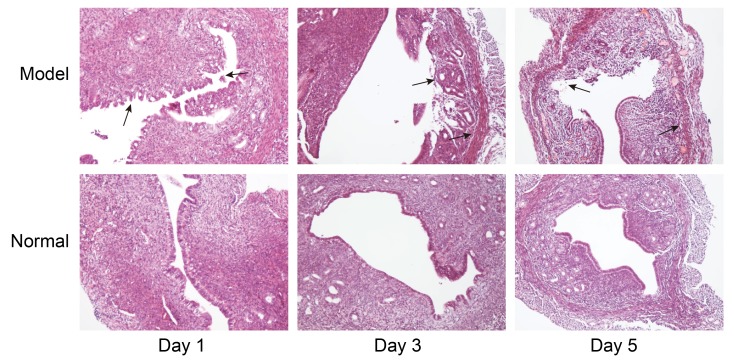
Figure 2.
Examination of endometrium in Model mice. Right uterine horns were collected from Model mice and examined for fibrosis by haematoxylin and eosin staining at days 1, 3 and 5 following endometrial damage; the left uterine horns were undamaged and used as a Normal control. Black arrow indicates endometrial fibrosis. Magnification, ×100.

