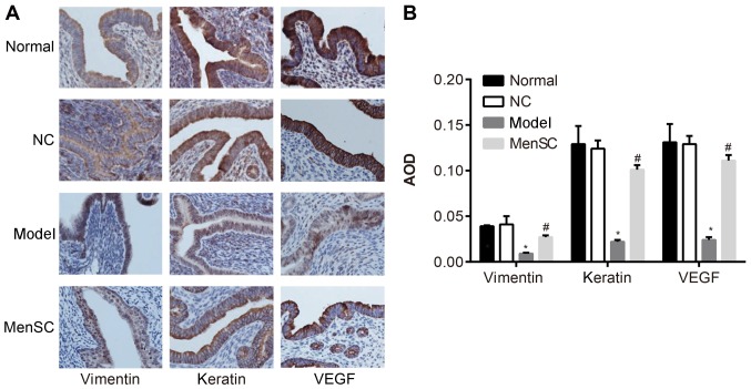
Figure 5.
Expression of vimentin, VEGF and keratin proteins in the endometrium of Model mice. (A) Immunohistochemical staining of endometrium from mice in each group; magnification, ×400. (B) Quantification of the expression of vimentin, VEGF and keratin from Part A. *P<0.05 vs. Normal; #P<0.05 vs. Model. AOD, average optical density; MenSCs, menstrual blood-derived stem cells; NC, negative control; VEGF, vascular endothelial growth factor.

