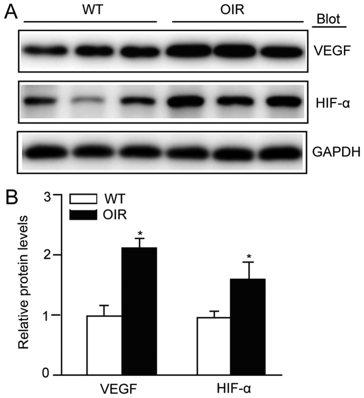
Figure 2.

VEGF-A expression is activated in the retinas of OIR mice. (A) Immunoblot analysis of the protein expression levels of VEGF-A and HIF-1α in the retinas. (B) Quantification revealed an increase in the expression levels of VEGF-A and HIF-1α in the retinas of the OIR mice compared with WT mice. The relative protein expression level was normalized to GAPDH (n=3 mice per group). Data are presented as the mean ± standard deviation of the mean. *P<0.05 vs. WT mice. OIR, oxygen-induced retinopathy; WT, wild-type; VEGF, vascular endothelial growth factor; HIF-1α, hypoxia inducible factor-1α.
