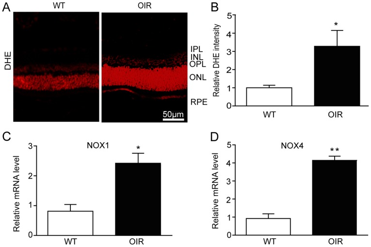
Figure 3.
Retinal oxidative stress is increased in OIR mice. (A) The reactive oxygen species levels in the retinas of the WT and OIR mice were detected using the DHE fluorescent probe and fluorescent images of the retinas were visualized with a fluorescence microscope. Scale bar, 50 µm. (B) Quantification of DHE intensity in the retinas (n=6 mice per group). The retinas were harvested for RNA extraction and qPCR detection. (C) NOX1 and (D) NOX4 were upregulated at the mRNA level in the retinas from the OIR mice compared with the WT mice. The relative mRNA expression levels were normalized to GAPDH (n=6 mice per group). Data are presented as the mean ± standard deviation of the mean. *P<0.05, **P<0.01 vs. WT mice. IPL, inner plexiform layer; INL, inner nuclear layer; OPL, outer plexiform layer; ONL, outer nuclear layer; RPE, retinal pigment epithelium; OIR, oxygen-induced retinopathy; WT, wild-type; VEGF, vascular endothelial growth factor; DHE, dihydroethidium.

