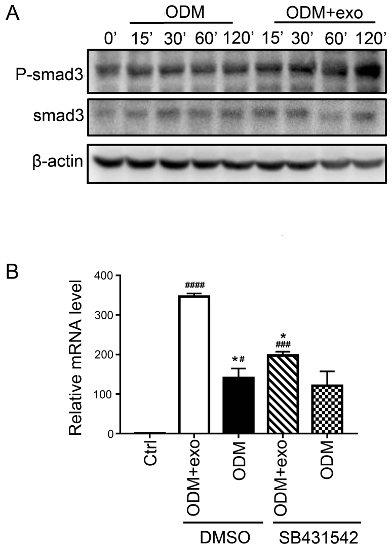
Figure 4.
3T3L1-exo activates 3T3L1 preadipocytes to undergo osteogenic differentiation via TGF-β signaling. (A) Western blot analysis detected p-smad3 and smad3 expression in 3T3L1 cells following stimulation with 3T3L1-exo for the indicated durations. (B) Reverse transcription-quantitative polymerase chain reaction was conducted to determine the mRNA expression levels of runt-related transcription factor 2 in 3T3L1 cells stimulated with ODM and/or 3T3L1 exo following treatment with the transforming growth factor-β inhibitor (SB431542). *P<0.05 vs. DMSO+ODM+exo; #P<0.05, ###P<0.001, ####P<0.0001 vs. control. (ODM + exo vs. ODM: DMSO, P=0.0204; SB431542, P=0.2204; Ctrl vs. DMSO+ODM+exo, P<0.0001; Ctrl vs. DMSO+ODM, P=0.0285; Ctrl vs. SB431542+ODM+exo, P=0.0009; Ctrl vs. SB431542+ODM, P=0.0883; DMSO+ODM+exo vs. SB431542+ODM+exo, P=0.0133; DMSO+ODM vs. SB431542+ODM, P=0.9662). Ctrl, control; DMSO, dimethyl sulfoxide; exo/3T3L1-exo, 3T3L1 cell derived-exosomes; ODM, osteogenic differentiation medium; P, phosphorylated; Smad3, SMAD family member 3.

