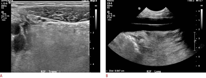
Fig. 1. A 13-year-old boy with acute appendicitis.
A. Grey-scale ultrasonography shows echogenic peri-appendiceal mesentery, which prompted a thorough examination of the right iliac fossa with a different transducer. B. Using the iliac vessels as an acoustic window deeper into the pelvis revealed an inflamed appendiceal tip (callipers).

