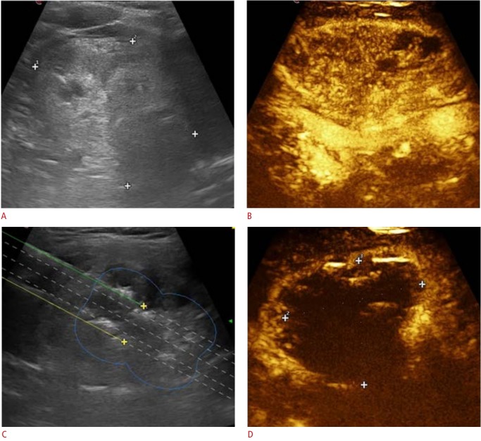
Fig. 1. Ultrasonography showing laser treatment of a 62-year-old patient with a benign thyroid nodule.
A. Ultrasonography scan before treatment demonstrates a large, isoechoic, non-homogeneous thyroid nodule. B. Contrast-enhanced ultrasonography of the same nodule before treatment shows intense enhancement of the nodule. C. Ultrasonography during treatment shows the insertion of two laser fibers with the use of dedicated planning software. Hyperechoic areas due to gas formation are seen around the needle tips (green and yellow lines, yellow markers, and blue circle line indicate expected fiber path, expected position of needle tip, and expected area of ablation with 1 pull-back, respectively). D. Contrast-enhanced ultrasonography performed after treatment demonstrates lack of enhancement in the treated area (markers).

