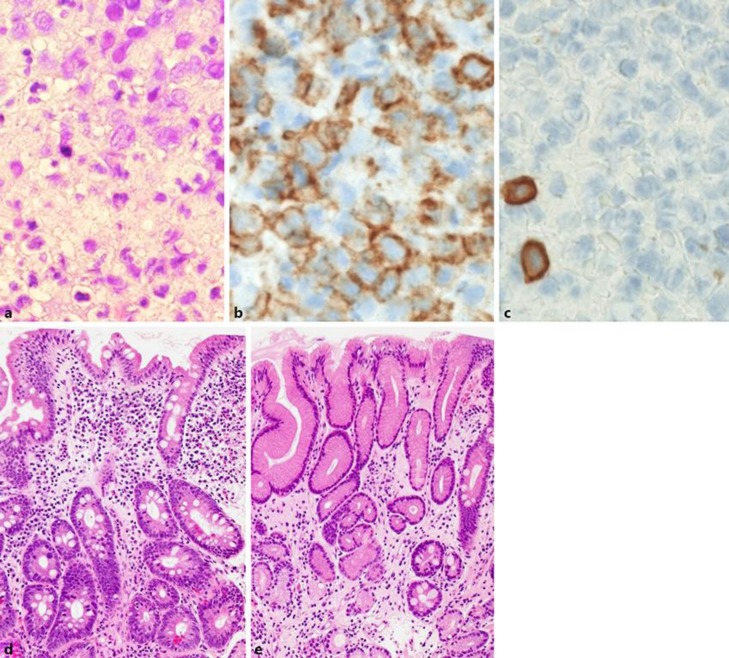
Fig. 2.
a Histology with HE staining at initial diagnosis (magnification ×400). b Positive staining of lymphoma cells by CD20 antibody in immunohistology (magnification ×600). c Negative staining of lymphoma cells by CD3 antibody in immunohistology. Only a few scattered normal T cells were positive (magnification ×600). d Histology with HE staining on the biopsy taken 12 months later after the initial diagnosis (magnification ×100). e Histology with HE staining in the biopsy tissue taken after H. pylori eradication treatment in 2018 (magnification ×100).

