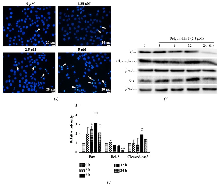
Figure 2.
Detection of PPI-induced cell apoptosis. Images of Hoechst 33258 staining were observed by fluorescence microscopy following treatment with PPI (1.25, 2.5, and 5 μM) for 12 h (magnification, x400), white arrows indicate apoptotic cells (a). HepG2 cells were incubated with PPI (2.5 μM) for 0, 3, 6, 12, and 24 h. The expression levels of apoptosis-associated proteins in HepG2 cells were detected by western blotting (b). The alterations in the expression levels of Bcl-2, Bax, and cleaved-caspase3 were statistically analyzed (c). Data were obtained from three independent experiments. The results were presented as the mean ± standard deviation. ∗p < 0.05 and ∗∗p < 0.01, vs. the control group (0 h). Bcl-2, B-cell lymphoma 2; Bax, Bcl-2-associated X; PPI, polyphyllin I.

