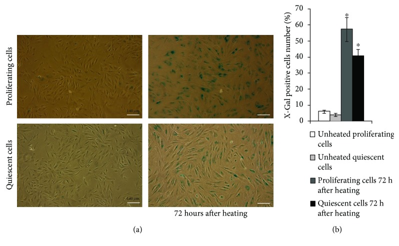
Figure 3.
HS induces appearance of SA-β-Gal-positive cells in both proliferating and quiescent cultures. (a) SA-β-Gal staining. The senescent cells were detected with SA-β-Gal staining kit Ob: 10x; scale bar = 140 μm. (b) Quantitative assay of SA-β-Gal-positive cells. At least 500 cells from different fields of view were analyzed. Data from three independent experiments are presented. ∗The difference vs unheated cells (t-test, p < 0.05).

