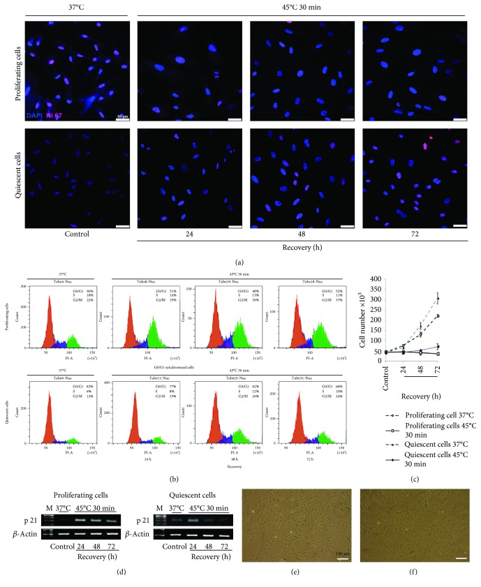
Figure 5.
Resumed proliferation of quiescent and proliferating cells after HS. (a) Immunofluorescence assay of proliferating and quiescent eMSC with anti-Ki-67 antibodies. Nuclei were contrasted with DAPI; scale bar = 50 μm. (b) FACS analyses of cell cycle of proliferating and quiescent eMSC after HS. (c) FACS analyses of cell viability of proliferating and quiescent eMSC after HS. (d) RT-PCR analysis of the p21 level in proliferating and quiescent cells. Data of three independent experiments are presented. (e, f) X-Gal staining. The progeny of HS-survived cells from proliferating (e) and quiescent (f) cultures. Ob: 10x; scale bar = 140 μm.

