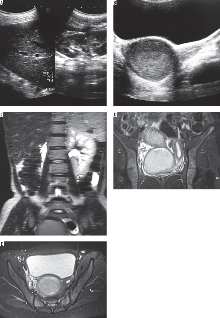
Figure 2.
A) Ultrasonographic (USG) image showing the empty right renal fossa and normal left kidney. B) Pelvic USG image showing the normal left uterine horn and dilated right horn with echogenic contents (hematometra). C) Coronal magnetic resonance image (MRI) showing the normal left kidney and absent right kidney. D) Coronal pelvic MRI showing dilated right uterine horn with haematometra and normal left horn. E) Axial MRI through the pelvis showing the normal left uterine horn with dilated right horn with haematometra

