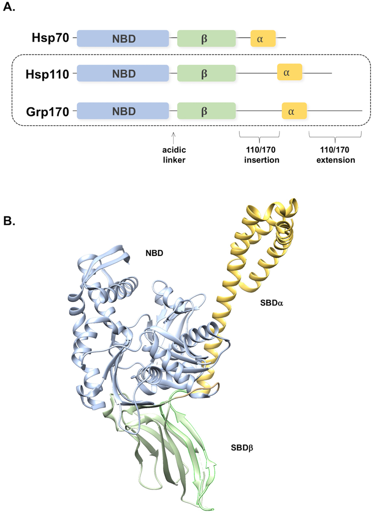
Figure 1. A, Domain architecture of Hsp70 superfamily.

NBD, nucleotide binding domain. β, β-sandwich peptide binding domain. α, α-helical bundle. Locations of additional sequences in the Hsp110 and Grp170 chaperone classes relative to Hsp70 are indicated, as is the acidic linker. B, Ribbon structure of the yeast Hsp110 Sse1 (PDB file 2QXL), domains colored as in A and labeled as indicated.
