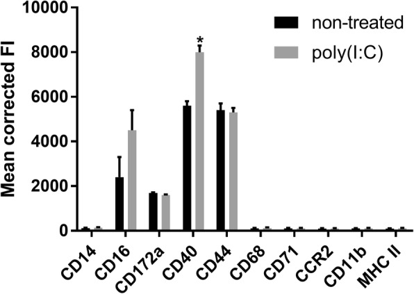
Fig. 1.

Flow cytometric analysis of Bomac cells. Bomac cells were labeled with antibodies against CD14, CD16, CD11b, CD172a, CD44, MHC II, CD40, CD68, CD71, CCR2 and using single color staining. Another set of cells were stimulated with p(I:C) as described in Material and Methods, and then similarly stained for flow cytometry. At least 100,000 events were acquired in FACS Calibur flow cytometer and analyzed in FlowJo software. Results are shown as mean fluorescence intensity (MFI). Six separate stainings for each group of cells were performed. * = p ≤ 0.05
