Figure 3.
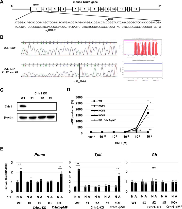
Characteristics of CRHR1-KO AtT-20 cells. (A) Target sequences of sgRNAs for CRISPR/Cas9 gene KO in Crhr1 gene exon 1. (B) Nucleotide sequences of part of Crhr1 exon 1 in WT AtT-20 cells and Crhr-KO AtT-20 cells (left). Schematic membrane topology of Crhr1 protein in WT AtT-20 cells and three Crhr1-KO AtT-20 clones obtained with TMHMM Server version 2.0 (right). (C) Representative Western blots for Crhr1 and β-actin in WT AtT-20 cells and three Crhr1-KO AtT-20 clones (#1, #2, and #3). (D) cAMP levels in WT AtT-20, three Crhr1-KO AtT-20 clones (KO#1, KO#2, KO#3), and Crhr1-KO AtT-20 cells transfected with Crhr1-pMF plasmid (KO+Crhr1-pMF) treated with increasing concentrations of CRH for 30 min. Relative values were calculated separately for each group based on the value of the group treated with 0 M CRH. Results are mean ±SD. *P < 0.05 vs WT; **P < 0.01 vs WT. (E) Quantitative RT-PCR analysis of Pomc, Tpit, and Gh gene expression in WT AtT-20, three clones of Crhr1-KO AtT-20 (#1, #2, #3), and Crhr1-KO AtT-20 cells transfected with Crhr1-pMF plasmid (KO+CRHR1-pMF) after 24-h treatment at normal pH 7.4 (N) and acidic pH 6.8 (A) medium. Results are mean ± SD. *P < 0.05 vs pH 7.4; **P < 0.01 vs pH 7.4. n.s., not significant.

