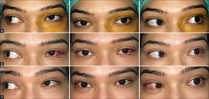
Figure 5.
(a) Preoperative photograph of a patient with left eye exo-DRS. The patient underwent supramaximal LR recession (15 mm) in the affected eye leading to the improvement in exodeviation but sill a residual exotropia was present (b) which was corrected by LR recession in the other eye leading to orthophoria (c)

