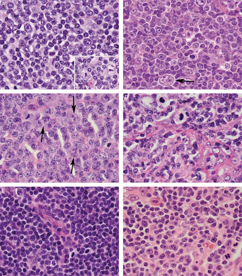
FIGURE 2.
Cytologic features of NLPHL with atypical T cells. Atypical T cells demonstrated a morphologic spectrum but were typically larger than the small lymphocytes within primary follicles (A, case 1; B, case 5; C, case 7; D, case 4; E, case 6; F, case 11), with vesicular (A) or dispersed (B-E) chromatin, irregular nuclei (B-D), occasional prominent nucleoli (B, F), moderately abundant pale eosinophilic cytoplasm (B-C, F), and increased mitotic activity (B-F). Rare scattered LP cells with multilobated nuclei, pale chromatin, and prominent nucleoli were visible within the T-cell-rich areas, both interspersed among the atypical T cells and at the periphery of primary follicles (A inset; B-C, arrows). In case 4, the T cells were associated with fine compartmentalizing background fibrosis (D).

