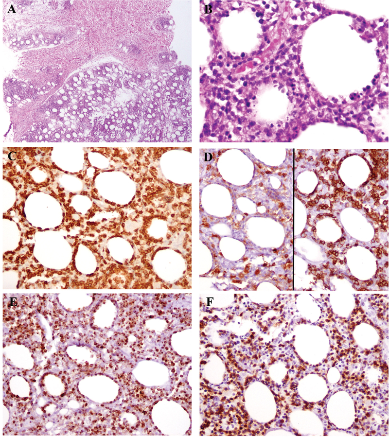
Fig. 1.

The findings from patient 4 illustrate those typically seen in pediatric SPTCL. The dermis was uninvolved (A, top), while a lobular panniculitic-like infiltrate was present in subcutaneous fat (A, bottom). Atypical lymphoid cells rim adipocytes, and karyorrhectic debris is abundant (B). The atypical lymphocytes express CD3 (C). They are negative for CD4 (D, left) and positive for CD8 (right). Granzyme B (E) and beta F1 (F) are also positive
