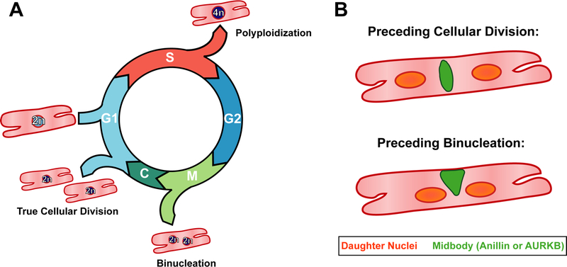
Figure 1:

A) The postnatal cardiomyocyte is capable of progressing through multiple cell cycle variants. If the CM exits following DNA synthesis, a process known as polyploidization, it remains mononucleated and contains a tetraploid (4n) nucleus. Furthermore, CMs can complete mitosis but not cytokinesis, leading to the formation of two diploid (2n) nuclei, known as binucleation. True cellular division, the goal of cardiac regenerative therapies, only occurs if the cardiomyocyte is able to fully complete cytokinesis, resulting in two daughter CMs.
B) Hesse et al demonstrate that there are two major criteria for differentiating cellular division and binucleation: 1. The midbody positioning, here depicted in green, differs between the two events. Specifically, the midbody is found to be located symmetrically between the two daughter nuclei if the cell will complete cytokinesis and divide, but asymmetrically if instead the cardiomyocyte will binucleated. The authors demonstrate the midbody can be visualized by either anillin or AURKB immunofluorescence. 2. The distance between the daughter nuclei varies, with the distance between the nuclei being greater preceding cellular division than binucleation.
