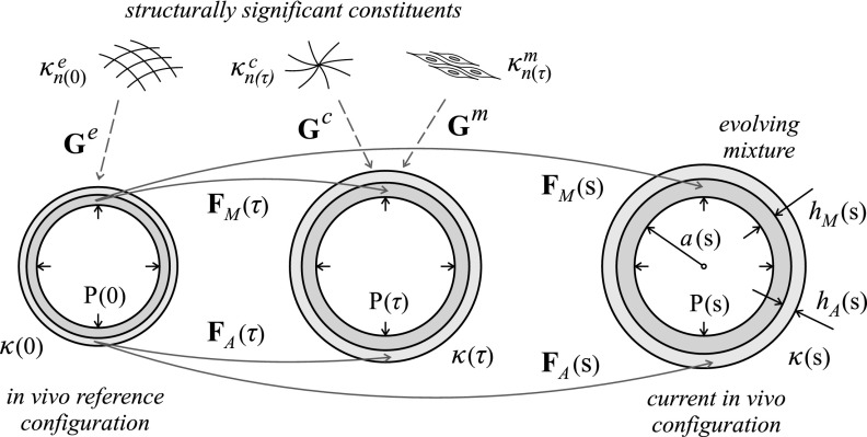
FIG. 6.
Evolving in vivo configurations of a bilayered arterial wall from time s = 0 (original homeostatic reference configuration κ(0) ≡ κo) to an arbitrary time s > 0 [current configuration κ(s)], showing the different deformation gradients FM and FA experienced by the medial and adventitial layers over time, in particular at times 0 ≤ τ ≤ s. Shown, also, are the different natural configurations for the different structurally significant constituents α (elastin “e,” smooth muscle “m,” and collagen “c”), which are deposited with separate but constant deposition stretches Gα within both layers at the indicated deposition times τ (except for the smooth muscle, present in media only), as well as geometric parameters a (luminal radius), hM (medial thickness), and hA (adventitial thickness) and luminal pressure P, which can also change over time.

