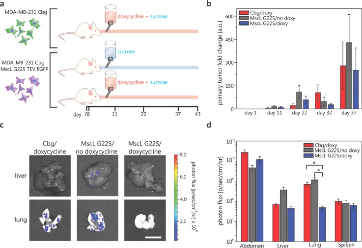
FIG. 1.
In vivo experiment for determining MscL's effect on cancer cell metastasis. (a) Cartoon description of in vivo experiments. MDA-MB-231 cells with doxycycline inducible expression of MscL G22S and constitutive luciferase expression and MDA cells with constitutive luciferase-only were injected under the mammary fat pad of immunodeficient mice on day 0. Three cohorts of mice were then studied: negative control group (1) mice with MDA-MB-231 MscL G22S luciferase cells with sucrose feed (n = 4), (2) mice with MDA-MB-231 luciferase only cells with doxycycline and sucrose feed (n = 5), and experimental group (3) mice with MDA-MB-231 MscL G22S luciferase cells with doxycycline and sucrose feed (n = 5). (b) Mean primary tumor size fold change at the site of initial injections as determined using bioluminescence imaging of mice on different days. Error bars represent the standard error of the mean. Differences in the total area-under-the-curve for bioluminescence do not differ among groups (p > 0.4). (c) Images of the extracted liver and lung with luminesce signal false coloring and the corresponding photon flux scale from a mouse of each cohort on day 43 relating to metastatic cancer cells at these secondary sites. Scale bar = 1 cm. The logarithmic plot of the average luminescence signal, the result of metastatic cancer cells, described as photon flux for various organs of each cohort. Error bars represent the standard error of the mean. The vertical axis starts above the luminescence background signal at 5 × 10⁶ p/s cm2 sr. Two-tailed student t-test of log transformed data: *p ≤ 0.01.

