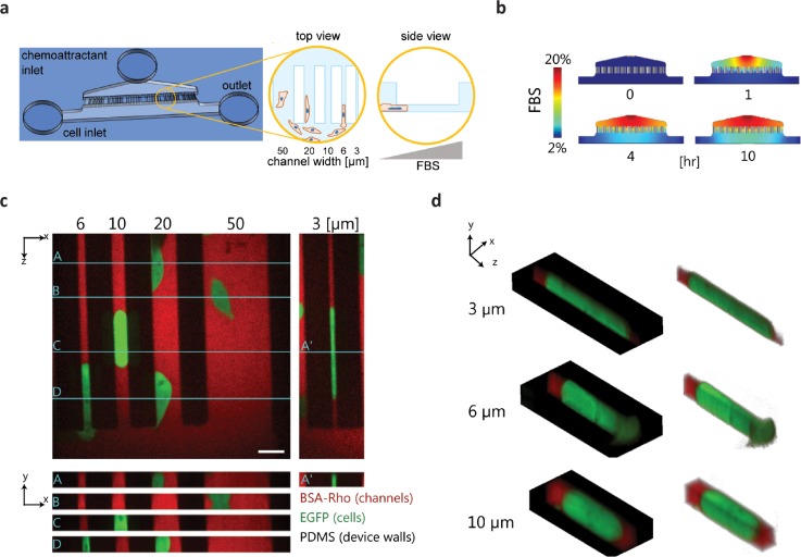
FIG. 4.
Microfluidic platform for studying cancer cell migration across 2D channels and narrow 3D constrictions. (a) Cartoon of the PDMS microfluidic migration device. Cells were added to the device via a cell inlet and flow into the bottom section of the device, adhered to the glass substrate, and migrated across channels of various sizes in response to an FBS chemoattractant gradient as shown in the zoomed-in views. (b) Images of COMSOL multi-physics simulation of the FBS gradient in the microfluidic device at different time points. (c) Fluorescence images showing orthogonal views, y-x and z-x, of EGFP expressing cells in the microfluidic device channels. The z-x cross-sections of the labeled, turquoise lines on the y-x view are shown for cells in all channel widths. The PDMS/device walls appear black in the images, and the channels were filled with media containing BSA-rhodamine. Scale bar = 20 μm. (d) Isometric view of the cells in the narrow 3D constriction channels.

