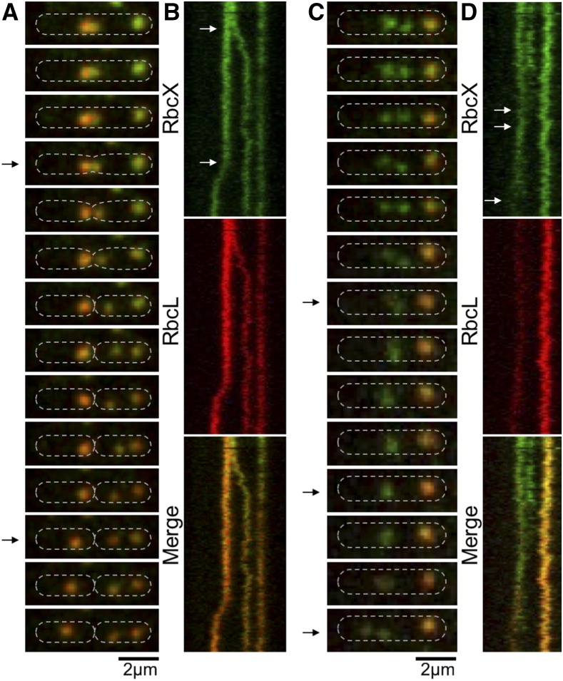
Figure 6.
Dynamics of RbcX-Rubisco assembly in live Syn7942 cells using time-lapse confocal fluorescence imaging. A, Time-lapse confocal images of a RbcX-eYFP/RbcL-CFP cell, showing the dynamic locations and interactions of RbcX and Rubisco during the carboxysome birth event and a cell-dividing process. Time interval, 1.25 min. B, Kymographs of RbcX-eYFP (green) and RbcL-CFP (red) assembly. Arrows indicate a carboxysome birth event and a cell-dividing event (bottom), as shown in A. Time interval, 1.25 min. Scale bar, 2 µm. C, Time-lapse confocal images showing the dynamic locations and interactions of RbcX and Rubisco during the fusion and splitting processes of RbcX-containing spots. Time interval, 1.25 min. D, Kymographs of RbcX-eYFP (green) and RbcL-CFP (red) assembly. Arrows indicate a fusion event and a dividing event (bottom) of RbcX-containing spots, as shown in (C). Time interval, 1.25 min. Scale bar, 2 µm.

