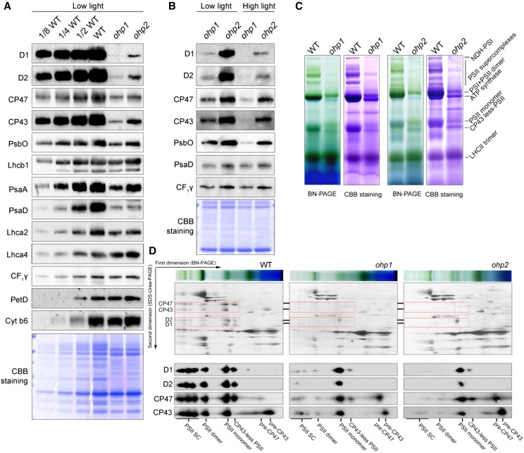
Figure 2.
Accumulation of thylakoid protein complexes in the ohp1 and ohp2 mutants. A, Immunoblot analysis of representative thylakoid proteins. Thylakoid membranes isolated from wild-type (WT), ohp1, and ohp2 plants were separated by 15% (w/v) SDS-urea-PAGE and then probed with antibodies as indicated. Proteins were loaded on an equal protein level. B, Immunoblot analysis of thylakoid proteins in the ohp1 and ohp2 mutants. The mutants grown under low-light conditions (50 μmol photons m−2 s−1) were shifted to high-light conditions (300 μmol photons m−2 s−1) for 2 d. Thylakoids were isolated for immunoblot analysis. Proteins were loaded on an equal protein level. C, BN-PAGE analysis of thylakoid protein complexes. Thylakoids corresponding to 10 µg of chlorophyll were solubilized with 1% (w/v) dodecyl-β-d-maltopyranoside and separated by 5% to 12% (w/v) BN-PAGE. The gels were stained with Coomassie Brilliant Blue (CBB) for visualization of the proteins. D, 2D BN/SDS-urea-PAGE analysis of the thylakoid membrane complexes. Thylakoid complexes were separated by BN-PAGE (C) and further subjected to 2D SDS-urea-PAGE. The gels were stained with Coomassie Brilliant Blue or probed with antibodies against D1, D2, CP43, and CP47. The positions of D1, D2, CP43, and CP47 in various PSII complexes are framed with red dotted boxes. SC, Supercomplexes.

