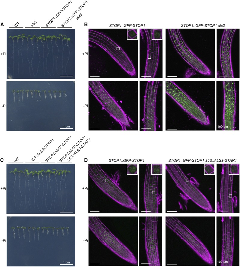
Figure 8.
Accumulation of STOP1 protein in the nucleus of root cells of different genotypes. A and C, Morphologies of 6-d-old seedlings of the various genotypes grown on +Pi and −Pi media. B, GFP signals of STOP1::GFP-STOP1 in roots of wild-type (WT) and als3 seedlings grown on +Pi and −Pi media. D, GFP signals of STOP1::GFP-STOP1 in roots of wild-type and 35S::ALS3-STAR1 seedlings. For the observation of GFP signals, seeds with different genotypes were germinated on +Pi medium and grown for 3 d. The seedlings then were transferred to +Pi or −Pi medium for 24 h before they were photographed. The cell walls were stained with propidium iodide (shown in magenta). The insets in B and D show closeup views of representative root cells. Bars = 1 cm (A and C) and 100 μm (B and D).

