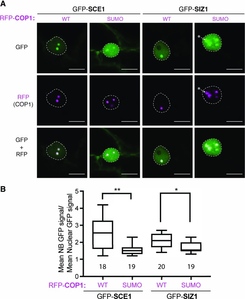
Figure 5.
Colocalization of GFP-tagged SCE1/SIZ1 with RFP-COP1 NBs is compromised following mutation of the COP1 SUMO acceptor site. A, Nuclear localization pattern of GFP-tagged SCE1 and SIZ1 in the presence of RFP-COP1 or the RFP-COP1SUMO SUMO acceptor site mutant (Lys-193Arg). * marks amorphous NBs in the GFP-SIZ1, RFP-COP1SUMO combination. Micrographs from top to bottom: GFP, RFP, and their merged signals. Scale bars, 10 µm; WT, wild type. B, Quantification of the average GFP signal intensity in the NBs per nucleus divided by the average fluorescence signal in the nucleus (with the data of three biological replicates pooled with at least five nuclei per replica). Total number of nuclei analyzed is shown. Significant differences were detected using an unpaired Student’s t test assuming unequal variances; **P < 0.01, *P < 0.05. Conditions were identical to those in Figure 1.

