Figure 1.
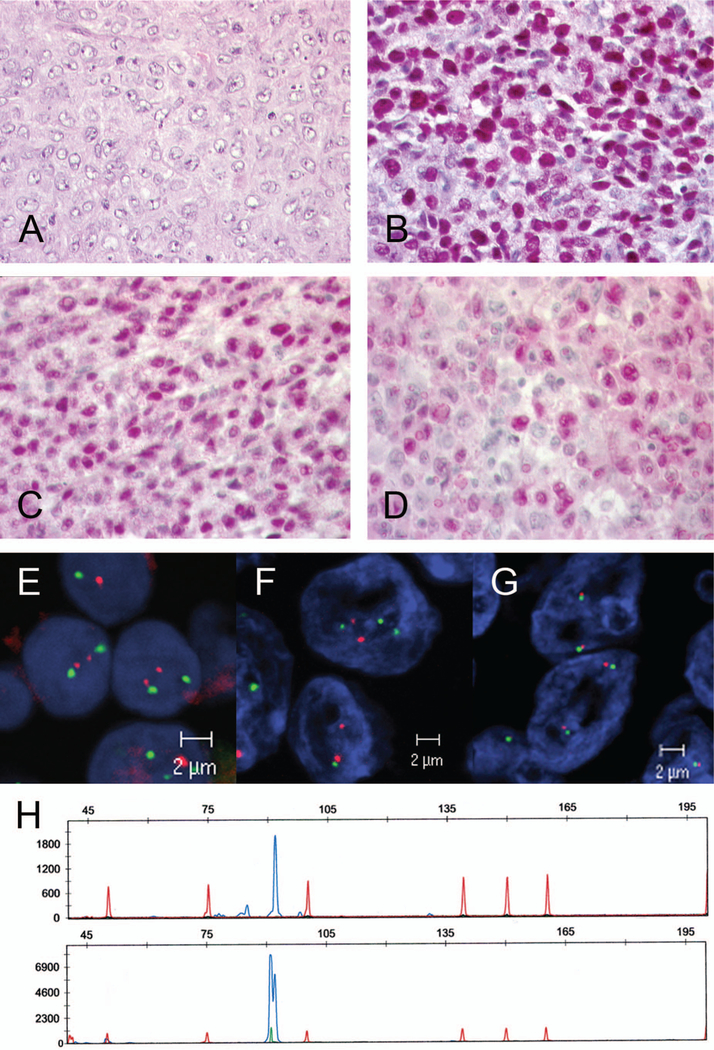
Case 17: Richter transformation of B-CLL. (A) Diffuse large B-cell lymphoma with centroblastic morphology (H&E, original magnification × 400). (B) MUM1 strong nuclear positivity in the majority of tumor cells (immunoperoxidase, original magnification × 400). (C) BCL6 positivity in the majority of tumor cells (immunoperoxidase, original magnification × 400). (D) Cyclin D1 expression in the tumor cells. Note that cyclin D1 was detectable in a majority of cells with weak to moderate intensity (original magnification × 400). (E, F) Interphase FISH analysis with dual color fusion probe for t(11;14)(q13)(q32). Note that neither B-CLL (E) nor transformed DLBCL (F) harbored a t(11;14) chromosomal translocation involving the CCND1 gene locus. (G) FISH analysis using a dual color break-apart probe for the MYC gene locus was not indicative for a chromosomal break involving the MYC gene locus. (H) Fragment length analysis of the CDR3 region of the immunoglobulin heavy chain gene revealed a monoclonal peak of the same size in B-CLL (upper panel) and in Richter transformation (lower panel), demonstrating that both tumors were clonally related.

