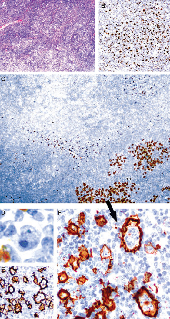
Figure 2.
Nodular lymphocyte-predominant Hodgkin lymphoma (NLPHL) with increased numbers of large cells (case 7). A, Areas of typical NLPHL with many singly scattered lymphocyte-predominant (LP) cells (inset) amidst a vaguely nodular background of lymphocytes and few histiocytes. B, Oct-2 stain highlights LP cells admixed with small B cells within nodules. C, Low-power view of CD15 stain showing numerous CD15+ LP cells in areas with increased numbers of large cells (**, bottom right) in contrast to adjacent areas of typical NLPHL clearly lacking CD15 expression (*, upper left). Only scattered granulocytes stain intensely in these latter areas. D, These cells are CD30) and strongly CD20+ (E). F, High-power view showing cytological atypia of the CD15+ large cells.

