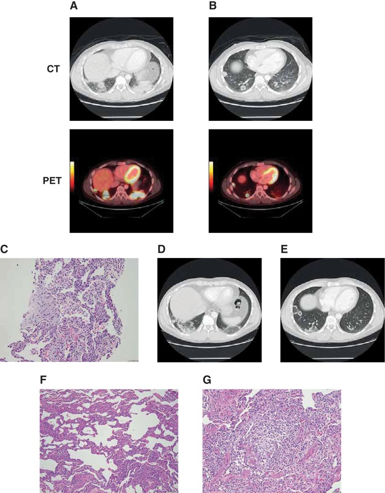
Figure 1.
Patient with pneumonitis on programmed death‐1 inhibitor therapy. Bilateral basilar lung consolidations (A) and pulmonary nodules, including cavitary nodules (B) detected on routine PET‐CT at cycle 28; cellular interstitial pneumonia and organizing pneumonia detected on interventional radiographic‐guided right lung biopsy (C); centrifugal expansion and central clearing of both the bilateral consolidations and pulmonary nodules on follow‐up CT (D–E); cellular and interstitial pneumonia (F) with multiple intra‐alveolar poorly formed granulomas and scattered eosinophils (G) identified on wedge resection of the involved right lower and upper lobe lung regions. Abbreviations: CT, computed tomography; PET, positron emission tomography.

