Fig. 5.
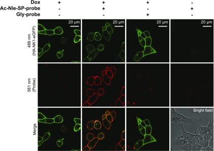
Ac-Nle-SP-probe binds to the NK1 receptor constructs at the cell surface. Dox-induced or uninduced Flp-In T-REx 293 cells harboring HA-NK1-eGFP were treated with Ac-Nle-SP-probe or Gly-probe (1 μM at 4°C for 1 hour) before exposure to UV light (15 minutes) and were then treated with DyLight 594-conjugated streptavidin before they were imaged using a confocal microscope. HA-NK1-eGFP and the DyLight 594–labeled probe were excited simultaneously at 488 nm (upper panels) and 561 nm (middle panels), respectively. Colocalization between the receptor and the probe was visualized (yellow color; lower panels). The bright field image demonstrates that cells were present in the uninduced −Dox panels.

