Fig. 6.
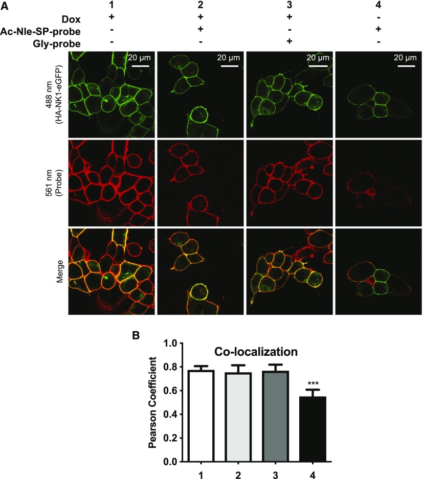
Ac-Nle-SP-probe covalently captures NK1 receptor constructs upon UV activation. (A) Dox-induced Flp-In T-REx 293 cells expressing HA-NK1-eGFP were treated with Ac-Nle-SP-probe (1 μM at 4°C for 1 hour) before they were activated with UV light for 15 minutes (samples 1 and 3) or kept in the dark (samples 2 and 4). Cells were then either treated immediately with DyLight 594–conjugated streptavidin (samples 1 to 2) or were first exposed to Ac-Nle-SP (10 μM at 4°C for 3 hours) (samples 3 to 4). They were then imaged using a confocal microscope. HA-NK1-eGFP and the DyLight 594–labeled probe were excited simultaneously at 488 nm (upper panels) and 561 nm (middle panels), respectively. Colocalization between the receptor and the probe was visualized (yellow color; lower panels). (B) Colocalization was quantified by generating green-red pixel intensity scatterplots for each pixel and determining the Pearson correlation coefficient for four representative images for each condition. Data are means + S.D. Significant statistical difference was determined using analysis of variance with Tukey’s multiple comparison test (***P ≤ 0.001).

