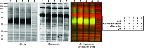
Fig. 7.
Detection of Ac-Nle-SP-probe NK1 receptor interactions. Dox-induced (lanes 1–4) or uninduced (lane 5) Flp-In T-REx 293 cells harboring HA-NK1-6xHis were treated with either Ac-Nle-SP-probe (lanes 2, 4, and 5) or Gly-probe (lane 3) and then exposed to UV light (lanes 1–3 and 5) or maintained in the dark (lane 4). Following cell lysis, membrane preparations were generated and resolved by SDS-PAGE. These were immunoblotted with an anti-6xHis antiserum (left panel) or streptavidin (center panel). Merging of these two images and pseudocolor labeling identifies the receptor-probe complex as yellow (right panel).

