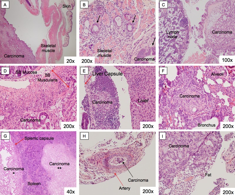
Figure 3. Histology of pancreatic cancer PDOX metastases.
Organs from PDOX mice with gross intra-abdominal metastases were harvested. There were metastatic deposits seen in the abdominal wall (A, B), within the peritoneum (C), small bowel (D), liver (E), lung (F), spleen (G), artery (H), and the retroperitoneum (I). These lesions retained the histoarchitecture of the primary tumor.

