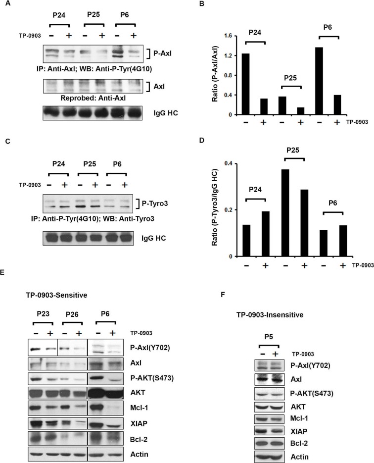
Figure 4. TP-0903 (tartrate salt) modulates CLL B-cell signaling proteins.
(A) TP-0903 inhibits tyrosine phosphorylation on Axl in CLL B-cells from ibrutinib treated cohort:Axl was immunoprecipitated from the DMSO or TP-0903 treated (20–24 hours) CLL B-cell lysates (n=3), followed by Western blot analysis using a global anti-phosphotyrosine (4G10) antibody. The blot was stripped and reprobed with an antibody to Axl. IgG HC was used as loading control. (B) Levels of P-Axl expression in CLL B-cells are quantified using ImageJ software and are represented as a ratio of P-Axl/Axl in the bar graph. (C) Effect of TP-0903 on Tyro3 phosphorylation: Total tyrosine phosphorylated proteins were immunoprecipitated from the DMSO or TP-0903 treated CLL B-cell lysates (n=3) used above using anti-phosphotyrosine antibody (4G10), followed by Western blot analysis using anti-Tyro3 antibody. IgG HC was used as loading control. (D) Levels of P-Tyro3 expression in CLL B-cells are quantified using ImageJ software and are represented as a ratio of P-Tyro3/IgG HC in the bar graph. (E) Impact of TP-0903 on Axl phosphorylation at kinase domain (Y702) and downstream signaling: DMSO or TP-0903–treated CLL B-cell lysates from TP-0903-sensitive patients (n=3) were analyzed for the status of phosphorylation at Axl (Y702) and AKT (S473) by Western blot analyses using specific antibodies. Respective blots were then stripped and reprobed with Axl or AKT antibody. TP-09003 treated same lysates were also used to determine the expression of anti-apoptotic proteins Mcl-1, XIAP, and Bcl-2 in Western blot using specific antibodies. Actin was used as a loading control. CLL patients (P23, P24; on ibrutinib treatment, P25, P26; scheduled for ibrutinib treatment and P6; progressed while on ibrutinib treatment) are indicated by arbitrary numbers. (F) Impact of TP-0903 on P-Axl (Y702), P-AKT (S473) and downstream signaling in CLL B-cells from a TP-0903-insensitive patient:DMSO or TP-0903 (0.50 μM)–treated CLL B-cell lysates from patient ‘P5’ were analyzed for the status of phosphorylation at Axl (Y702) and AKT (S473) by Western blot analyses using specific antibodies. Respective blots were then stripped and reprobed with Axl or AKT antibody. TP-09003 treated same lysates were also used to determine the expression of anti-apoptotic proteins Mcl-1, XIAP, and Bcl-2 by Western blot using specific antibodies. Actin was used as a loading control. CLL patient (P5) is indicated by arbitrary number.

