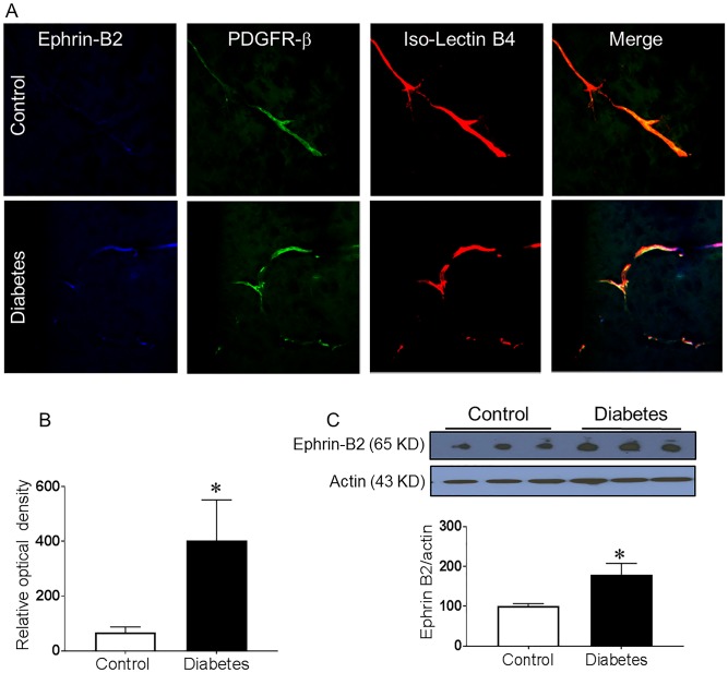
Fig 1. Increased Ephrin-B2 expression in pericytes in diabetes.
Four weeks male Wistar rats were injected with low dose streptozotocin followed by 8-weeks of high-fat diet (HFD, 45% fat). Brains were isolated and fixed. Brain sections were reacted with anti-Ephrin-B2 antibody and Iso-Lectin-B4. (A) Co-localized Ephrin-B2 (Blue), PDGFR-β (green) on Iso-Lectin-B4 (red) was compared between diabetic and control Wistar rats. Diabetic rats showed increased Ephrin-B2 expression in perivascular area that was co-localized with the pericytes marker. (B) Quantification of the relative optical density. (C) Western blot analysis showed a significant increase in Ephrin-B2 expression in the brain cortex of diabetic rats compared to control. (N = 4–5, *P<0.05 vs control).

