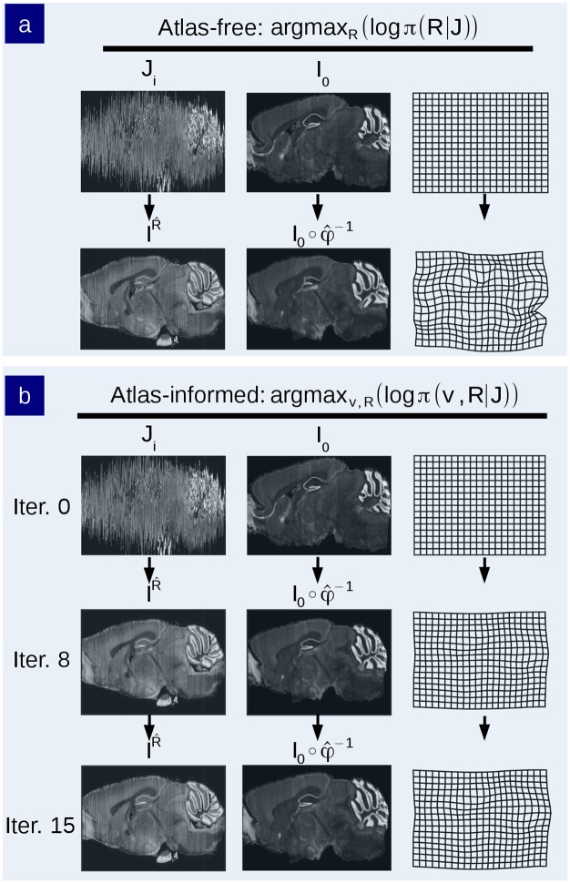
Fig 8. Comparison of reconstruction and mapping using atlas-free and atlas-informed models on data from the MBAP database.
a) Reconstruction of an MBA Nissl-stained brain histological stack using the atlas-free method. Top row shows the histological stack and Allen mouse brain atlas. Bottom row shows the reconstructed histological stack alongside the deformed phantom atlas I, and the diffeomorphic change of coordinates . b) Reconstruction using the atlas-informed method. Top row shows the histological stack and Allen mouse brain atlas. Middle row depicts intermediate iterations of the reconstructed stack alongside the deformed atlas and coordinate grid. Bottom row shows the convergence point of algorithm.

