
Fig. 2. Visualization of chromosome translocations by time-lapse microscopy.
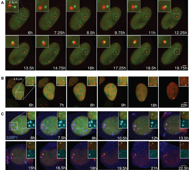
(A and B) Time-lapse microscopy of NIH3T3duo cells transfected with IScel. Maximal projected image sequences of representative movies show pairing of (A) proximal and (B) distal DSBs. Scale bars, 5 μm.(C) Formation of LacO (green)-TetO (red) translocation relative ot repair foci in NIH3T3 duo cells stably expressing BFP-53BFP1 (blue). Scale bar, 5 μm.